Volume 1 Issue 8, August 2014

Editorial
Gupta AS

Advanced Rhabdomyosarcoma During Pregnancy
Zarariya A, Katke RD, Lewis P, Agrawal GD.

Fetal Nasal Agenesis
Bakre T, Gupta AS.

Ichthyosis Uteri
Sathe PA, Patil LY, Vaideeswar P, Mayadeo NM.

Endometrial Osseous Metaplasia
Fernandes G, Patil A, Samant PY, Parulekar SV.

Near - Miss Mortality In Pregancy With Coarctation Of Aorta
Dudhe M, Saxena N, Khadkikar R, Qureshi S, Chauhan AR.

Focal Spontaneous Colpocleisis Leading To Apareunia
Saraogi MR, Kiran U.

Post Hysterectomy Vault Calcification – Therapeutic dilemma
Valvi D, Rashmi Prasad R, Parulekar SV.

Genesis of True Broad Ligament Leiomyoma
Parulekar SV

Editorial

Gupta AS

Merriam- Webster dictionary defines 'atresia' as the absence or closure of a natural passage of the body. Vaginal atresia can be congenital or acquired. Congenital occurs from birth though it is usually diagnosed after puberty. Acquired occurs in an adult woman who has previously menstruated or even borne children.
Acquired atresia may involve the entire length of the vagina or may be partial. Closure of the previously patent vagina may occur at its introital end, some distance beyond the introitus or at the upper end near the fornices though usually the presence of the cervix prevents a complete obliteration of the upper end of the vagina. The vaginal normally remains in the closed state with its anterior and the posterior wall apposed to each other mainly in its middle and the lower segment.
Vaginal is lined by squamous epithelium which matures and thickens under the influence of estrogens. It becomes rich in glycogen. Doderleins bacilli produce lactic acid in the vagina. This lowers the vaginal pH. The thick vaginal epithelium and the acidic pH are the defence mechanisms of the vagina. Vaginal mucosa is thin in the prepubertal and in the post menopausal women. In lactating women also there may be a relative deficiency of the estrogen hormone making the vagina thin and less acidic. Some women on antiestrogens like GnRh antagonists, SERMS without hormonal 'add-back' also develop estrogen deficiency. Women receiving radiotherapy, chemotherapy, or who have undergone oophorectomy also have estrogen deficiency. Estrogen deficiency is known to result in cytological changes in the vagina. Fragmentation of elastin, proliferation of its connective tissue and hyalinization of its collagen fibers are all well documented. These changes at the cellular level results in injury to the epithelium, abrasions, granulation tissue formation, erosions and ulcerations. While the woman leads a physically active sexual life the vaginal patency is maintained but in estrogen deficient women who are sexually inactive the vaginal walls remain apposed to each other. Presence of inflammation, erosions or granulation tissue in the vagina epithelium then predisposes to adhesion formation and when these are dense then the vagina gets obliterated. In some cultures women after childbirth stay away from their spouse and the resumption of sexual activity is delayed. Dyspareunia is the usual presenting symptoms in such patients. They then seek medical aid. Sometimes a pin hole opening may remain and this permits the menstrual flow whenever menstruation resumes like that in a lactating woman. Diagnosis can be made on clinical examination with the use of a speculum or a gloved finger. If the lower part of the vagina is obliterated separation of the labia may show the completely or partially closed introitus. Attempt to insert a speculum or a digit will result in pain and the tissues will feel firm and rigid. Shortening of the vagina can also be diagnosed when the upper part of the vagina is obliterated. Treatment can be combined medical, surgical and life style adaptation. In case of dense peri introital adhesions estrogen replacement in the form of local creams or oral supplementation can be attempted. However, with dense adhesions surgical approach may also be required as seen in the case report by Dr. Sarogi M.R. However, in their patient they have not tried the use of estrogen creams. After surgical correction the patient should use estrogen for healing and resume an active sexual lofe or use vaginal dilators to prevent the recurrence of vaginal atresia. Encouraging these women to continue active sexual life is a very important life style modification especially in post menopausal women where long term estrogen deficiency exists.
I present the July issue of our journal and hope our esteemed readers gain useful insight of the various clinical cases.

Advanced Rhabdomyosarcoma During Pregnancy

Author Information

Ashish Zarariya*, Rajshree Dayanand.Katke**,Preeti Lewis***, Grishma.D.Agrawal****
(*Associate Professor, ** Medical Superintendant & Associate Professor, *** Assistant Professor, **** Third Year Resident. Department of Obstetrics and Gynaecology, Cama And Albless Hospitals, Grant Government Medical College, Mumbai, India.)

Abstract

We report an interesting case of advanced rhabdomyosarcoma (RMS) in a teenage pregnancy leading to mortality. A 19 year old married girl presented with 8 months of amenorrhea and a wart like perianal lesion. She was lost follow up for a month and came in emergency in critical condition with septicaemia, hyperkalemia and acute renal failure (ARF). The wart sized lesion had progressed in a month to gross perianal mass which was extending inside pelvis up to the lower lumbar region. The patient succumbed within 8 hours of admission. On post-mortem histopathological examination, the lesion was diagnosed as a rhabdomyosarcoma.

Introduction

A case of rhabdomyosarcoma with near term pregnancy is a exceedingly rare event. Cancer in pregnancy itself is relatively rare. Most frequently reported sites are breast, head and neck, lymphomas and melenomas.[1] The origin of RMS is from tissue that imitates normal striated muscle.[2] Common sites are head and neck, genitourinary tract, thorax and abdomen. RMS is classified by international classification as embryonal, botryoid, spindle cell, alveolar, and undifferentiated. Out of this the botryoid and spindle cell types, considered to be the subtypes of embroyonal RMS, occur in 0-10 age group and have superior prognosis.[3] Alveolar RMS has poor prognosis, and its incidence is evenly distributed in 0-19 age group.[4]
RMS is classified by international classification as Embryonal and Alveolar.
Out of this Botryoid and Spindle cell, considered to be the subtypes of Embroyonal.[3]

Case report

A 19 year old Primigravida Unregistered, came to ANC OPD for Registration  with 7 months amenorrhea and perianal wart like growth.


Figure 1. Perianal lesion at the first visit.

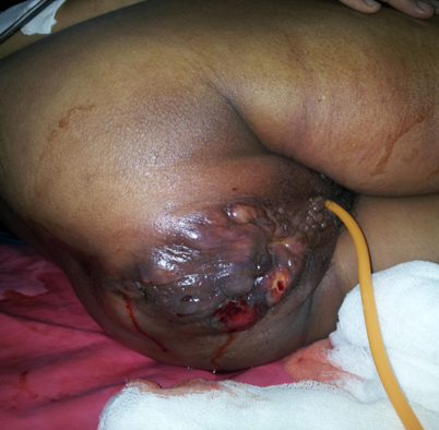
Later after 1 month, the patient presented in a critical state in emergency,  with palpable 3-4 cm hard lump in the left breast, edema in both lower limb up to thighs and indurated ulcerative growth with multiple grouped vesicles present around anus. The growth was extending inside the pelvis with gross left inguinal lymphadenopathy.


Figure 2. Perianal lesion at the second visit.

She had an intrauterine fetal death at 34-36 weeks. On per vaginal examination, the cervix could not felt due to the growth. She was treated with supportive line of management. Her investigations were suggestive of hyperkalemia, septicemia and ARF. She died within 8 hours of admission. Her postmortem examination showed a palpable lymph node in left inguinal region 3 cm in diameter, swelling of the left breast with palpable lymph node 2 cm diameter, a 10x8 cm whitish colored growth in the left lower lumbar region and left side of the pelvis. It extended to the perianal region in the form of nodules. The uterus showed features of acute myometritis with degenerated decidua.


Figure 3: Postmortem abdominal gross findings.

Histopathological examination of the tumor mass around the vertebral column showed a high grade malignant tumor with small round tumor cells with rhabdoid differentiation- a rhadomyosarcoma. A part of uterus showed infiltration and abscess formation, acute myometritis and degenerated decidua.

Discussion

The incidence of RMS is very low. Due to its rarity and diagnostic diversity, very little is known about the etiology of RMS. Several environmental factors have increased risk of developing RMS, such as  paternal cigarette smoking,[5] advanced maternal age and x-ray exposure in utero,[6] maternal and child’s antibiotic use,[7] stillbirths[8] and maternal recreational drug use[9]. In addition genetic changes may also play an important role in RMS development. Familial syndromes associated with inherited gene defects, like Li-Fraumeni syndrome and neurofibromatosis, have been associated with RMS.[10] RMS relative 5-year survival rates have not increased significantly over the past 30 years; RMS has one of the worst prognosis with high rates of mortalities. The diagnosis of a rhabdomyosarcoma depends on recognition of differentiation of its cells toward skeletal muscle cells. Immunohistochemical marker of rhabdomyosarcoma are MyoD1 and Myogenin. In our case immunohistochemistry could not be done as it was a post-mortem case and the facility was not available in our institute. Pertaining to our case a diagnosis of malignancy must be kept in mind for a painful ulcerative growth in this age group.Our patient presented as a teenage pregnancy and so the tumor was all the more rapidly progressive in nature leading to catastrophic, life threatening events ultimately resulting in untimely mortality of the patient.

References

1.        Donegan Wl: Cancer and pregnancy. CA Cancer J Clin 1983;33:194-214.
2.        Stout AP. Rhabdomyosarcoma of the Skeletal Muscles. Ann Surg. 1946;123:447–472.
3.        Gurney JG, Young JL, Roffers SD, Smith MA, Bunin GR. SEER Pediatric Monograph. National Cancer Institute; 2005. Soft Tissue Sarcomas.
4.        Ries LAG, Smith MA, Gurney JG, Linet M, Tamra T, Young JL, Bunin GR, editors. Cancer incidence and survival among children and adolescents: United States SEER Program 1975–1995. Bethesda: National Cancer Institute;1999.
5.        Gurney JG, Severson RK, Davis S, et al.: Incidence of cancer in children in the United States. Sex-, race-, and 1-year age-specific rates by histologic type. Cancer 1995;75(8): 2186-95.
6.        Ries LA, Kosary CL, Hankey BF, et al., eds.: SEER Cancer Statistics Review, 1973-1996. Bethesda, Md: National Cancer Institute, 1999. 
7.        Grufferman S, Wang HH, DeLong ER, Kimm SY, Delzell ES, Falletta JM. Environmental factors in the etiology of rhabdomyosarcoma in childhood. J Natl Cancer Inst 1982;68:107–113.
8.        Grufferman S, Gula MJ, Olshan AF, Falletta JM, Pendergrass TW, Buckley J, Maurer HM. In utero x-ray exposure and risk of childhood rhabdomyosarcoma. Paediatr Perinat Epidemiol 1991;5:A6.
9.        Hartley AL, Birch JM, McKinney PA, Teare MD, Blair V, Carrette J, Mann JR, Draper GJ, Stiller CA, Johnston HE, et al. The Inter-Regional Epidemiological Study of Childhood Cancer (IRESCC): case control study of children with bone and soft tissue sarcomas. Br J Cancer 1988;58:838–842.
10.    10. Ghali MH, Yoo KY, Flannery JT, Dubrow R. Association between childhood rhabdomyosarcoma and maternal history of stillbirths. Int J Cancer 1992;50:365–368.

Citation

Zarariya A, Katke RD, Lewis P, Agrawal GD. Advanced Rhabdomyosarcoma during pregnancy. JPGO 2014 Volume 1 Number 8 Available from: http://www.jpgo.org/2014/08/advanced-rhabdomyosarcoma-during.html

Fetal Nasal Agenesis

Author Information

Tejashree Bakre*, Gupta AS**
(* Third Year Resident, ** Professor. Department of Obstetrics and Gynecology, Seth G.S. Medical College and K.E.M Hospital, Mumbai, India.)

Abstract

A well controlled, pregnant, diabetic patient with bad obstetric history was diagnosed with fetus having single umbilical artery (SUA) and absent nasal bone. Quadruple marker test revealed low risk for trisomy 21, 13, 18 and neural tube defects. Amniocentesis was done and fluid was sent for karyotyping and FISH. FISH revealed no aneuploidy of chromosomes 13, 18, 21 and sex chromosomes. Karyotype revealed paracentric inversion of chromosome 7.

Introduction

Inversions account for about 10% of structural chromosomal aberrations, with pericentric inversions being more common than paracentric rearrangements probably due to diagnostic difficulties in the latter group.  Pericenteric inversions and most frquently associated with  chromosome 2 . Incidence reported is around 11% . Paracenteric inversions  are frequently seen in chromosomes 7 and 3  (19% and 16%, respectively)[1]

Case Report

      A 27 year old Gravida 3 with 2 spontaneous abortions and  38.3 weeks of pregnancy, a known case of diabetes mellitus on lifestyle modifications for the last 1 year was referred to our hospital casualty on 1st April 2014 in view of severe pre eclampsia. There were no pre monitory symptoms. BP on admission was 180/110 mmHg, there was mild pallor, bilateral pedal edema, knee jerks were normal, chest was clear,  uterus was full term, relaxed with fetus in vertex presentation and regular fetal heart sounds.  On vaginal examination cervical os was 1.5cm dilated, poorly effaced,  vertex presentation,  station -3, membranes were present.  Urine albumin was nil. Patient was diagnosed with gestational hypertension at 31 weeks at BP of 146/90 mm Hg and was started on T. Labetalol 100mg qid which was modified to 100mg bid 4 weeks later. Serological, biochemical and hematological investigations were done. Tab Nifedipine 10 mmg 6 hourly was started. Injection Magnesium sulphate was started by Pritchard regime and induction of labour was done after all reports were found to be normal. HbA1c in the second month of pregnancy was 6.3% suggesting good control of sugars. She was evaluated in private for bad obstetric history for previous two spontaneous first trimester abortions. TORCH titres were negative for IgM antibodies. Lupus anticoagulant and anti cardiolipin antibodies both IgM and IgG were negative. Thrombophilia profile was within normal limits. A malformation scan done at 21 weeks gestation revealed an absent nose and a single umbilical artery. A quadruple marker test was done in view of single umbilical artery which revealed low risk for trisomy 21, trisomy 18 and neural tube defects.  Subsequently amniocentesis was done and amniotic fluid was sent for karyotyping and FISH tests. No aneuploidy was detected for chromosomes 13, 18, 21 and sex chromosomes on FISH. This was done on interphase nuclei.


Figure 1: FISH Report.

Karyotyping was done. It diagnosed paracentric inversion of chromosome 7. This was a report written on her case records. Patient did not have the mapped karyotype document with her. Parental karyotyping was done.  It revealed normal karyotype. She had uneventful antenatal course till term. She delivered a female child of 2.075 kg weight, by face to pubis vaginal delivery. It was a fresh still birth. Baby had absent nose, hypertelorism, laryngeal stenosis and single umbilical artery.


Figure 2: Baby showing Facial Dysmorphia. N: indicates the absent nose. Hypertelorism (widely spaced eyes) is clearly seen.

Discussion

Statistics suggest that the nasal bone can be absent in normal foetuses (chromosome component normal) in 3% of the cases with an absent nose. In two thirds of fetuses with trisomy 21 the nasal bone is absent.[2] Single umbilical artery is a risk factor for structural and chromosomal  abnormalities.[3] One third of the foetuses have structural abnormalities and about 10% foetuses have chromosomal anomalies (aneuploidy) like trisomy 13, and 18. Trisomy 21 is not found to be associated with a single umbilical artery (SUA).[4]
When sonography diagnosed the absent nasal bone and SUA the patient was evaluated by a quadruple marker test to determine the risk of trisomy 21, 18 and neural tube defects. An amniocentesis was done as the quadruple test returned a low risk result for aneuploidy. A quadruple test is not recommended as per existing scientific evidence in such cases.[5] Karyotyping is recommended when a patient with single umbilical artery also shows intrauterine growth retardation, or any structural abnormality.[5] The amniotic fluid obtained by amniocentesis was sent for karyotyping and FISH. Karyotyping diagnosed a  paracentric inversion of chromosome 7. FISH was normal.
Paracentric inversion of chromosome 7 can have varied clinical presentations. Syndromes like Michelin tire , Williams-Beuren, ectrodactyly-ectodermal dysplasia-cleft syndromes have been described in literature in association with paracentric inversion of chromosome 7.  In all of these syndromes there is facial dysmorphia, developmental and behavioural disorders In "Michelin tire syndrome" there is hirsutism, smooth muscle hamartoma, facial dysmorphia, submucous cleft palate, genital and dental anomalies, seizures and moderate developmental delay. But in our case only few of these features were noted. A peculiar feature of this syndrome is that the baby and mother have apparently identical paracentric inversions of the long arm of chromosome 7.[6] However in our case parental karyotyping was normal. Normal parental karyotyping is commonly associated in cases of congenital absence of nose as these cases are usually sporadic.  Literature describes two cases with absent nose involved chromosome 9 anomalies, and in a third case there was de novo reciprocal translocation between chromosomes 3q and 12p.[7] However chromosome 7 anomalies have not been reported to be associated with congenital absence of nose.
Antenatal diagnosis of total arrhinia based on ultrasonographic findings has been described. Arhinia per se is not life threatening but the immediate and long-term outcomes of the neonate are dependent on associated anomalies. The afflicted neonate  may need emergency resuscitation due to respiratory distress. In this case there was laryngeal stenosis which was the probable cause of death. The technique and timing of the reparative procedures of the nasal structures is also a medical controversy. Parental counselling is important. A multidisciplinary team effort is required for best neonatal results.[7]

References
  1. Muss B and  Schwanitz G: Characterization of Inversions as a Type of Structural Chromosome Aberration: International Journal of  Human Genetics, 200;7(2):141-161.
  2. Cicero S, Longo D, Rembouskos G : Absent nasal bone at 11–14 weeks of gestation and chromosomal defects : Ultrasound Obstet Gynecol 2003;22: 31–35
  3. Onofriescu M, Nemescu D, Martiniuc V: 21st World Congress on Ultrasound in Obstetrics and Gynecology  Oral poster abstracts OP04.06  Prenatal echographic diagnosis of single umbilical artery Ultrasound in Obstetrics & Gynecology 2011;38 (Suppl 1):56–167.
  4. Geipel A , Germer U, Welp T: Prenatal diagnosis of single umbilical artery: determination of the absent side, associated anomalies, Doppler findings and perinatal outcome : Ultrasound in Obstetrics & Gynecology: 2000;15(2),114–117.
  5. Nyberg D, Nyberg B, McKenna C : : 21st World Congress on Ultrasound in Obstetrics and Gynecology  Oral poster abstracts OP04.04 Absent or near absent nasal bone in the 2nd trimester :Ultrasound in Obstetrics & Gynecology 2011; 38 (Suppl. 1): 56–167.
  6. Schnur R,  Herzberg A,  Spinner N : Variability in the Michelin tire syndrome. A child with multiple anomalies, smooth muscle hamartoma, and familial paracentric inversion of chromosome 7q. : Journal of the American Academy of Dermatology 03/1993; 28(2 Pt 2):364-70.
  7. Merino A, De Perdigo A, Nombalais F, Yvinec M, Le Roux MG, Bellec V. Prenatal diagnosis of trisomy 9 mosaicism: two new cases. Prenat Diagn. 1993 Oct; 13(10):1001-7.
Citation

Bakre T, Gupta AS. Fetal Nasal Agenesis. JPGO 2014 Volume 1 Number 8. Available from: http://www.jpgo.org/2014/08/author-information-tejashree-bakre.html

Ichthyosis Uteri

Author Information

Sathe Pragati A*, Patil Lalita Y**, Vaideeswar Pradeep***, Mayadeo Niranjan M****
(* Associate Professor, ** Assistant Professor,  ***Additional Professor, Department of Pathology, **** Professor, Department of Obstetrics and Gynecology, Seth GS Medical College & KEM Hospital, Mumbai, India.)

Abstract

Ichthyosis uteri, a rare condition, is called so when the entire surface of the endometrium is replaced by keratinized stratified squamous epithelium. It is a benign lesion and its association with benign and malignant conditions has been reported in the literature. If widespread keratinization of endometrial surface is detected in the curettage or biopsy then an advanced examination should be done to rule out an underlying malignancy.

Introduction

The term ichthyosis uteri is used when there is widespread replacement of the surface endometrium by keratinized stratified squamous epithelium.[1] It is considered a benign lesion but its association with malignancy has been reported in the literature. [2] We report a case of ichthyosis uteri detected on endometrial curettage in an elderly postmenopausal woman.

Case Report

A 60-year-old woman, gravida 9, para 9 with eight living issues came with complaints of foul smelling white discharge of three months duration. Per vaginal examination showed a normal sized anteverted uterus. Bilateral fornices were free. On per speculum examination, foul smelling cervical discharge was noted. The vagina was normal. No growth was appreciated in the cervix. Ultrasonography of the pelvis showed 3x2x1.5 centimeters sized collection inside the uterine cavity. Both ovaries were normal. Clinical impression was endometritis with pyometra. The hematological and routine biochemical investigations were within normal range. The patient underwent cervical dilatation along with drainage of 50 ml of profuse greenish foul smelling purulent discharge from the uterine cavity. Papanicolau stain of the cervical discharge showed mature keratinized squamous epithelial cells on a background of neutrophils. Fractional endometrial curettage was performed after six weeks and the material was sent for histopathological examination. The microscopy showed prominent neutrophilic infiltrate with necrosis. Bathing in this exudates, were seen strips of keratinized squamous epithelium. The normal endometrial glands were covered by this epithelium (Figure 1a). There was low grade dysplasia of the squamous epithelium (Fig 1b). There was no evidence of associated endometrial malignancy. A diagnosis of ichthyosis uteri was given. The patient was administered oral antibiotics and discharged with an advice for a hysterectomy. She refused further treatment and was lost to follow-up, despite explanation of the associated risks.


Figure 1. Ichthyosis Uteri. 1a. Keratinized squamous epithelium with endometrial glands beneath. (Hematoxylin and Eosin, 100x); 1b. Low grade dysplasia seen in the squamous epithelium. (Hematoxylin and Eosin, 400x)

Discussion

First coined by Zeller in 1885, the term ‘ichthyosis uteri’ means widespread keratinization of the endometrium. [3] It is rarer compared to squamous metaplasia. Chronic trauma, repair, irritation, inflammation, foreign material, and estrogenic effects have all been implicated.[3] The etiology of endometrial keratinization is not well understood. This rare pathology has been seen in association with benign conditions like tuberculous endometritis, puerperal endometritis, endometrial polyps, hyperplasias, squamous papilloma and with pyometra as a result of cervical stenosis.[1,3] Hence, when ichthyosis is seen on endometrial curettage, one has to look for associated benign pathologies especially tuberculosis, endometrial polyps and hyperplasia.
According to some investigators, ichthyosis lacks malignant potential.[1] However, dysplastic and anaplastic changes in the squamous epithelium have been reported, which may predispose to   the rare endometrial squamous carcinoma in postmenopausal women.[1,2]
The main differential diagnosis to be considered before making a diagnosis of pure ichthyosis is the extension of squamous carcinoma of the cervix into the endometrial cavity. The primary tumor in such a case would have an infiltrating rather than a polypoid morphology. The extension of well differentiated squamous carcinoma from the cervix can be distinguished from ichthyosis by detailed examination of the lower genital tract, by the presence of koilocytic changes and the presence of dysplastic changes in the squamous epithelium which favors a diagnosis of squamous carcinoma extension from cervix. [1,3]
In our case, the predisposing factor seems to be repeated episodes of pyometra which is suggested by strips of squamous epithelium embedded within abundant neutrophilic exudates as seen on endometrial curettage. The low grade dysplasia seen in the epithelium could suggest the likelihood of associated squamous carcinoma cervix. Unfortunately, our patient declined any further treatment and hence the cause of ichthyosis could not be ascertained
To conclude, ichthyosis is a benign condition but can lead to or be associated with endometrial malignancy, hence the need for timely recognition of this condition.[2] The clinical diagnosis in such cases is most frequently pyometra which has to be thoroughly investigated, especially when it occurs in postmenopausal women. If widespread keratinization of endometrial surface is detected in the curettage or biopsy then an advanced examination should be done to rule out an underlying malignancy.[1]

References

1.      Fadare O. Dysplastic Ichthyosis uteri-like changes of the entire endometrium associated with a squamous cell carcinoma of the uterine cervix. Diagn Pathol 2006;1:8-11.
2.      Bagga PK, Jaswal TS, Datta U, Mahajan NC. Primary endometrial squamous cell carcinoma with extensive squamous metaplasia and dysplasia. Indian J Pathol Microbiol 2008;51:267-8.
3.      Bewtra C, Xie QM, Hunter WJ, Jurgensen W.Ichthyosis uteri: a case report and review of literature. Arch Pathol Lab Med 2005;129:e124-e125.

Citation

Sathe PA, Patil LY, Vaideeswar P, Mayadeo NM. Ichthyosis Uteri. JPGO 2014 Volume 1 Number 8. Available from: http://www.jpgo.org/2014/08/ichthyosis-uteri.html

Endometrial Osseous Metaplasia

Author Information
Gwendolyn Fernandes*, Asmita Patil**, PY Samant*** SV Parulekar****
(* Associate Professor, Department of Pathology, ** Senior Resident, *** Additional Professor **** Professor and Head, Department of Obstetrics & Gynecology, Seth GS Medical College & KEM Hospital, Parel, Mumbai, India.)

Abstract

Osseous metaplasia of the endometrium is a rare condition characterized by the presence of mature or immature bone in the endometrium. Most cases present with secondary infertility following an abortion or chronic endometritis, some patients are asymptomatic, while others have menstrual irregularities or menorrhagia. We present two cases of osseous metaplasia of the endometrium.

Introduction

Osseous metaplasia of the endometrium is a rare condition characterized by the presence of mature or immature bone in the endometrium. Most cases present with secondary infertility following an abortion or chronic endometritis, some patients are asymptomatic, while others have menstrual irregularities or menorrhagia. Ultrasound examination showing characteristic hyperechogenic pattern of osseous tissue within the uterus helps suspect the diagnosis. The final diagnosis is confirmed by hysteroscopy and removal of the bony tissue by curettage. Complete removal of the bony spicules from the endometrial cavity by hysteroscopy under ultrasonic guidance usually cures the patient.

Case Report 1

A 24 year old woman presented with secondary infertility for 8 years. She had had a spontaneous abortion at 4 months of amenorrhea 8 years ago, at which time she had undergone a blunt curettage. Since then she had had menorrhagia, the bleeding lasting for 8 to 10 days every 30 days. Her general and systemic examination revealed no abnormality. Her uterus was of 6 weeks' size, smooth and firm. There was no pelvic tenderness or mass. Her hemogram, blood sugars, liver and renal function tests, HIV, VDRL and her husband's semen analysis reports were within normal limits. Difficulty was encountered during passage of a uterine sound and bony spicules were seen in the endometrium during hysteroscopy. All the bony spicules were removed by a sharp curette under laparoscopic control to prevent uterine perforation. She made an uneventful recovery. Her menorrhagia was cured. Three months later she was lost to follow up, and her fertility status remains unknown.


Figure 1 – Microphotograph showing mature bone surrounded by blood clot, fibrinous material and inflammatory cells.


Figure 2 – Microphotograph showing higher magnification of the bony tissue.


Figure 3 – Microphotograph showing well-formed mature bone, calcific material and inflammatory cells.

Case Report 2

A 35 year old para 4 MTP 1 presented with abnormal uterine bleeding (irregular menses with soakage of 2 pads per day) for 4 years. She had had 4 normal deliveries followed by a medical termination of pregnancy at 3 months of amenorrhea 4 years ago. Her general and systemic examination revealed no abnormality. Her uterus was of normal size, smooth and firm. There was no pelvic tenderness or mass. Her hemogram, blood sugars, liver and renal function tests, chest radiograph and electrocardiogram were normal. Endometrial aspiration showed no malignant cells. Her ultrasonography showed a 2.9 cm sized calcified lesion, which was interpreted as either endometrial calcification or calcified submucosal leiomyoma. During dilatation and curettage, there was difficulty in passage of a uterine sound. Presence of bony spicules in the endometrium was suspected. These spicules were removed held by a long, curved, hemostat, followed by curettage. She made an uneventful recovery. Her menorrhagia was cured without any additional treatment.


Figure 4 -  Microphotograph showing calcified bone and inflammatory cells.


Figure 5 – Microphotograph showing abundant calcific material and mature bone.


Figure 6 – Microphotograph showing higher power view of the mature bone.

Discussion

Osseous metaplasia of the endometrium is a rare condition characterized by the presence of mature or immature bone in the endometrium.[1] Its estimated incidence is 3/10000, there being about eighty cases described in the literature.[2] It has been referred to by various names ectopic intrauterine bone, heterotopic intrauterine bone, endometrial ossification etc.[3]
Clinically the patients are in the reproductive age group. A history of a previous pregnancy or abortion has been reported in more than 80% cases.[,3,4,5] The interval between the antecedent pregnancy and detection of endometrial ossification varies from 2 months to 14 years.[6] In our cases, it was 8 and 4 years respectively. Most cases present with secondary infertility following an abortion or chronic endometritis, some patients are asymptomatic, while others have menstrual irregularities or menorrhagia.[5,7] One of our patients had secondary infertility and the other had menorrhagia. Ultrasound examination showing characteristic hyperechogenic pattern of osseous tissue within the uterus helps suspect the diagnosis. The final diagnosis is confirmed by hysteroscopy and removal of the bony tissue by curettage.
There have been various controversies regarding the etiology and pathogenesis of osseous metaplasia of the endometrium. There have been debates on whether the osseous tissue was of maternal or fetal origin. However genetic analysis of the osseous tissue, and comparison with the DNA of both the parents, have shown that there is no male paternal genetic material in it, ruling out a fetal origin. There have been many theories of its origin, such as dystrophic calcifications and ossification of post-abortive endometritis, heterotopia, metaplasia in healing tissue, metastatic calcification, and prolonged estrogenic therapy after abortion.[3,4,5,8,9]  The most accepted theory of the origin of the osseous tissue is metaplasia of endometrial stromal cells into osteoblastic cells which produce the bone.[6] Inflammatory conditions like endometrial tuberculosis, chronic endometritis, and pyometra, trauma of curettage or instrumentation are causes of chronic inflammatory pathology and these can result in endometrial osseous metaplasia.[6] In India, endometrial tuberculosis should be ruled out as a cause of infertility as well as endometrial calcification and ossification.
It is also important for pathologists to avoid making an erroneous diagnosis of malignant mullerian tumor on histology.[5,6,7] This nonneoplastic etiology should not be missed.
Osseous metaplasia is a treatable condition. Complete removal of the bony spicules from the endometrial cavity by hysteroscopy under ultrasonic guidance usually cures the patient.[6,10,11]

References

1.        Umashankar T, Patted S, RS Handigund RS. Endometrial osseous metaplasia: Clinicopathological study of a case and literature review. J Hum Reprod Sci 2010;3(2): 102–104.
2.        Kishore kumar BN, Dr. Deepak Das D, Shivaraj HG. Endometrial osseous metaplasia :  A case report. Int J Biol Med Res. 2012;3(2): 1865-1866.
3.        Hsu C. Endometrial ossification. Br J Obstet Gynaecol. 1975;82:836–9.
4.        Dutt S. Endometrial ossification associated with secondary infertility. Br J Obstet Gynaecol 1978;85:787–9.
5.        Bhatia NN, Hoshiko MG. Uterine osseous metaplasia. Obstet Gynecol 1982;60:256–9.
6.        Bahçeci M, Demirel LC. Osseous metaplasia of the endometrium: a rare cause of infertility and its hysteroscopic management. Hum Reprod 1996;11:2537–9.
7.        Shimizu M, Nakayama M. Endometrial ossification in a postmenopausal woman. J Clin Pathol 1997;50:171–2.
8.        Acharya U, Pinion SB, Parkin DE, Hamilton MP. Osseous metaplasia of the endometrium treated by hysteroscopic resection. Br J Obstet Gynaecol 1993;100:391–2.
9.        Waxman M, Moussouris HF. Endometrial ossification following an abortion. Am J Obstet Gynecol 1978;130:587–8.
10.    Coccia ME, Becattini C, Bracco GL, Scarselli G. Ultrasound-guided hysteroscopic management of endometrial osseous metaplasia. Ultrasound Obstet Gynecol. 1996;8:134–6.
11.    Lee JY, Lee HA, Kwon HM, Na SH, Hwang JY, Lee DH. A case of endometrial osseous metaplasia treated by hysteroscopic operation. Korean J Obstet Gynecol 2012;55(5):361-365.

Citation

Fernandes G, Patil A, Samant PY, Parulekar SV Endometrial Osseous Metaplasia. JPGO 2014 Volume 1 Number 8. Available from:  http://www.jpgo.org/2014/08/endometrial-osseous-metaplasia.html

Near - Miss Mortality In Pregnancy With Coarctation Of Aorta

Author Information

Monali Dudhe*, Neha Saxena*, Rashmi Khadkikar**, Shabnam Qureshi**, A. R. Chauhan***
(* Third Year Resident, ** Assistant Professor, *** Additional Professor. Department of Obstetrics and Gynecology, Seth GS Medical College and KEM Hospital, Mumbai, India.)

Abstract

Coarctation of aorta (stenosis commonly located at the junction of the arch of aorta and proximal descending aorta) may be diagnosed for the first time during pregnancy, as most patients are asymptomatic. Dilatation and dissection of the aorta can lead to increased maternal mortality; significant stenosis is a contraindication to pregnancy. However, successful pregnancies have been reported in women with uncorrected coarctation if preconception risk stratification is done. We report a primigravida who presented with chronic hypertension and breathlessness where the diagnosis of coarctation was made incidentally during active labor in view of differences in blood pressure (BP) readings of both arms. She underwent emergency lower segment cesarean section (LSCS) and remained stable postoperatively.

Introduction

Coarctation of aorta accounts for 5 to 8 % of patients with congenital heart disease.[1] Marked increase in maternal cardiac output and oxygen consumption in pregnancy is associated with left ventricular failure, aortic dissection or rupture, endocarditis and cerebrovascular accidents. Individual risk stratification depends upon the size of the aorta, presence of aneurysm, severity of the coarctation and presence of hypertension.

Case Report

A 23 year old primigravida with 38 weeks’ of gestation was referred to our tertiary centre for intensive care in view of breathlessness and fever with chills for 3 days. On examination, she had fever, tachycardia (pulse of 136/minute), tachypnea (respiratory rate of 40/minute) and hypertension - blood pressure (BP) of 150/80 mm Hg. On chest auscultation, air entry was reduced in the left lower zone. She had been diagnosed as chronic hypertension at 20 weeks of gestation and was on treatment with tablet methyldopa 250 mg three times a day. She had scoliosis since birth.
All routine hematological investigations were normal; however arterial blood gases revealed pH of 7.3 with oxygen saturation of 91%. Chest radiograph showed patchy opacity in left lower zone suggestive of pneumonia. Chest sonography showed minimal right sided pleural effusion with underlying consolidation; hence a provisional diagnosis of community acquired pneumonia was made. Cardiac cause of hypoxia was not suspected at this time. Obstetric ultrasonography showed severe oligohydramnios.
The patient was started on intravenous antibiotics and oxygen by mask as she was able to maintain her saturation only with 100% oxygen. Antihypertensives were continued. On day 2 of admission, she was transferred to intensive respiratory care unit in view of decreasing oxygen saturation, where she went into spontaneous labor.  At this time, in view of differences in BP readings of both arms, right upper limb BP 150/90 mm Hg and left upper limb BP 130/90 mm of Hg, emergency cardiology opinion was taken and 2- D echocardiography was done, which diagnosed severe coarctation of aorta just proximal to the left subclavian artery: diameter of aorta at coarctation site was 6 mm (compared to 14 mm at diaphragm), with peak systolic gradient of 100 mm of Hg across the descending aorta and  ejection fraction of 50%. Infective endocarditis prophylaxis was started and emergency LSCS was performed under epidural anesthesia, which was difficult due to scoliosis. Male baby weighing 2.76 kg was delivered with Apgar score of 9/10; liquor amnii was thick meconium stained. The patient was shifted to intensive cardiac unit postoperatively, where her course was uneventful. She was started on tablet metoprolol 12.5 mg daily, and discharged after ten days. CT aortogram postpartum showed hypoplastic aortic arch, as seen in figures 1 and 2. Aortoplasty is planned 6 months postpartum.


Figure 1: CT aortogram (AP and lateral) showing coarctation in lateral view.


Figure 2: Magnified view of coarctation of aorta.

Discussion

The vascular malformation in coarctation is a defect in the medial layer, giving rise to a prominent posterior infolding or “posterior shelf”. Proliferation of elastic tissue in the intima occurs distal to the coarctation causing intimal dissection or aneurysm formation.[2]
Coarctation may be associated with other cardiovascular anomalies like bicuspid aortic valve, left-sided obstructive or hypoplastic defects and ventricular septal defects, in almost 50% of patients. Extracardiac nonvascular anomalies of head and neck, skeletal, genitourinary, gastrointestinal, or respiratory systems may be present, as in our patient who had thoracic spine scoliosis. Intracranial aneurysm of circle of Willis is seen in 3-5% cases.
Aortic coarctation causes congestive heart failure in early life. In adults, it is usually asymptomatic; however, hypertension, headache, nosebleed, leg cramps, claudication, muscle weakness or neurologic changes may be seen. Clinical examination is diagnostic: systemic hypertension is almost invariable with differences in upper and lower extremity arterial pulses and BP; diminished and delayed pulses distal to the obstruction are characteristic. Hence it should be a routine practice in every patient with systemic arterial hypertension to simultaneously palpate brachial and femoral pulses to search for the “brachial-femoral delay” of significant aortic coarctation. Supine bilateral arm (brachial artery) BP and prone right or left supine leg (popliteal artery) BP should be measured to search for differential pressures.
In pregnancy, increased blood volume and increased estrogen levels contribute to hypertension and increased shear stress on aortic wall; systemic hypertension is hence the most common risk associated with patients of coarctation, and is responsible for dilatation and dissection of the aorta. Poorly controlled hypertension also leads to adverse neonatal outcomes like growth retardation, abruption and premature delivery. Antihypertensive therapy can cause hypotension distal to the coarctation site, leading to reduction in uteroplacental perfusion.[3] 
In unrepaired coarctation, as also in repaired coarctation with arterial hypertension, residual coarctation, or aortic aneurysm, the risk to the fetus and mother is increased, and maternal mortality rate may be as high as 3-8%. Risk of aortic dissection, rupture of cerebral aneurysm, congestive heart failure and angina pectoris are increased in late pregnancy, intrapartum and within 6 weeks postpartum due to hemodynamic and hormonal changes.
Conservative management is preferred in antenatal period. Control of hypertension by beta-blockers, management of congestive cardiac failure by digoxin and afterload reduction by diuretics should be done as per individual case assessment. Indications for surgical intervention include significant coarctation or re-coarctation with long standing hypertension, hemodynamically significant aortic stenosis and female patient contemplating pregnancy.
Percutaneous intervention is possible during pregnancy. It should be performed only if there is persistence of severe resistant hypertension or presence of maternal compromise; as there is high chance of dissection during procedure.[4] Surgical techniques include balloon dilatation with or without stent placement, patch aortoplasty with Dacron patch, and direct surgical repair with excision of the para-coarctation tissue.
LSCS was previously recommended due to greater risk of rupture of both aortic and cerebral artery aneurysms in late pregnancy, labor and early puerperium following vaginal delivery. However recent European Society of Cardiology (ESC) guidelines suggest vaginal delivery under epidural anesthesia is safe in both uncorrected and corrected coarctation.[4]
To conclude, coarctation was fortunately but incidentally diagnosed when our patient was in labor. Value of measuring BP in different limbs cannot be over emphasised in every patient of hypertension. Contribution of physician, cardiologist and anesthetist was important in this case, and possibility of aortic dissection and subsequent mortality was averted.

References

1.      Yavuz C, Soydinc HE, Tekbaş G and Karahan O. Pregnancy Complicated with Severe Recurrent Aortic Coarctation: Case Rep Vasc Med 2012; Article ID 865035. Available from: http://www.hindawi.com/journals/crivam/2012/865035/
2.      Wald RM, Sermer M, and Colman JM. Pregnancy in young women with congenital heart disease: Lesion-specific considerations. Paediatr Child Health. 2011;16:e33-e37.
3.      Connolly HM. Pregnancy in women with coarctation of the thoracic aorta. ACC Curr J Rev 1997;55:6-7.
4.      Warnes CA, Williams RG, Bashore TM, Child JS, Connolly HM, Dearani JA, et al. ESC Guidelines on the management of cardiovascular diseases during pregnancy: 2011;32:3147–3197.

Citation

Dudhe M, Saxena N, Khadkikar R, Qureshi S, Chauhan AR. Near - Miss Mortality In Pregancy With Coarctation Of Aorta. JPGO 2014 Volume 1 Number 8. Available from: http://www.jpgo.org/2014/08/near-miss-mortality-in-pregancy-with.html

Focal Spontaneous Colpocleisis Leading To Apareunia

Author Information

Mohit R Saraogi*, Usha Kiran**
(* Assistant Professor, ** Associate Professor **, Department of Obstetrics & Gynaecology, Cama & Albless Hospital, JJ Group of Hospitals, Mumbai, India.)

Abstract

Colpocleisis or obliteration of the vagina is a rare clinical entity with most cases being acquired in the form of Le Forts repair. Sponataneous Colpocleisis following a normal vaginal delivery is an unknown  phenomenon in medical history. Here we present to you a case report of a young woman who presented to us with spontaneous colpocleisis following a normal vaginal delivery at home with no precipitating or predisposing factors.

Introduction

Colpocleisis or obliteration of the vagina is derived from the greek term "kolpos" which refers to a fold in the greek tunic and "cleisis" which stands for occlusion or closure.[1] Therapeutic colpocleisis in the form of Le Forts repair, has long been accepted as a treatment modality for prolapse in elderly women who are unfit for major surgery or general anaesthesia.[2]  However spontaneous colpocleisis following a vaginal delivery is an unknown clinical entity with no documented literature available on its occurrence and management. The condition can be traumatizing for a young women in the reproductive age group as it can present with apareunia, dyspareunia, menstrual irregularities, hematometra, pyometra and endometriosis. We present the case report of a young woman who presented to us with spontaneous colpocleisis following a normal vaginal delivery at home.

Case Report

A 25 year old para 2 living 2 woman presented to our Out Patient Department (OPD) with complaints of apareunia following a vaginal delivery 1 year ago. The patient had undergone an uneventful cesarean section in her first pregnancy 5 years ago in view of non progress of labor. In her second pregnancy, the patient went into spontaneous labor 1 year ago and had a home delivery under the supervision of a dai (traditional midwife).
On direct questioning the duration of labor in the patient was approximately 12 hours. The delivery was uneventful and the patient was examined in a nearby municipal hospital immediately postpartum and later discharged without any intervention. There was no history of vaginal lacerations, postpartum hemorrhage or need for intervention in the patient after delivery. The patient gave history sexual abstinence for six weeks following delivery. On attempting to resume coitus, she had apareunia which had persisted till date. On direct questioning she also gave history of hypomenorrhea since 8 months with minimal soakage of 1 pad per day for 5 days. Her prior menstrual history was normal. There was no history of any other medical or surgical illness in the patient.
On examining the patient, her vital parameters were stable and her general and systemic examination were normal. On abdominal examination, her abdomen was soft.  Local examination showed no signs of chronic perineal tear. Per vaginal or per speculum examination was tried unsuccessfully as there was complete obliteration of the vaginal cavity with thick mucosal adhesive band across. A decision for examination under anesthesia with vaginal refashioning was taken for the patient.
      The patient was examined under spinal anesthesia in the operation theatre and the OPD findings were confirmed. A decision to proceed with vaginal refashioning was taken. A Foley’s catheter was introduced into the bladder so as to minimize the risk of bladder or urethral injury. A per rectal examination revealed that the uterus and the cervix could be felt above the level of the adhesions through the posterior vaginal wall. A small pin point dimpling was seen in the center of the vagina with a thin mucosal aperture of 1 mm, when the fleshy adhesive band was stretched using the finger in the rectum. The aperture would not admit a uterine sound.  A tuboplasty probe was passed through the aperture followed by a uterine sound. Then a series of progressively increasing Hegar’s dilators (No. 3 onwards) were passed through the vaginal opening to enlarge the lumen until a one finger digital examination could be performed. The digital examination showed that there were thick fleshy mucosal adhesions across the anterior, posterior and lateral vaginal walls in the lower end of the vagina. However the upper two thirds of the vagina was free of adhesions and the uterus and cervix could be digitally palpated and also visualized using a small sized speculum by stretching the adhesions slightly. This fleshy band of adhesions were lysed with a transverse incision and dissected fully using sharp scissors, taking care not to injure the bladder, urethra or the rectum. At the end of the surgery, the vagina admitted two fingers and a speculum examination could be done easily. Raw vaginal mucosa was sutured perpendicular to the line of incision and a vaginal Foley’s catheter was placed in the middle and lower one third of the vagina and inflated with 50 ml normal saline, so as to prevent contractures. This catheter was deflated and reinflated once a day. The patient was given broad spectrum antibiotics (cefotaxime and metronidazole) for 5 days and kept catheterized (bladder) for 24 hours. The patient was given Seitz bath 3-4 times a day and encouraged to maintain genital hygiene. A speculum and digital examination was repeated 72 hours after the surgery and the wound was found to be healing well. The vaginal Foley’s catheter was changed after 72 hours and removed after 1 week. The patient was discharged on day 7 of surgery and asked to follow up weekly on an OPD basis.
            On her first post operative visit, a digital examination was done. The vagina could admit two fingers and a speculum examination revealed a healthy cervix and vagina. Her second post operative visit confirmed the same findings, however there was an inflammatory ridge on the posterior vaginal wall made prominent by the contraction of the levator ani as a reflex action to pain.  The patient was anxious and complained of pain.  Stronger pain killers were prescribed to the patient along with lignocaine jelly for local application. As the wound had healed well she was asked to use vaginal dilators to prevent recurrence of adhesions and to enlarge the lumen. On her third postoperative visit, she was comfortable using dilators and the pain had reduced considerably. She was asked to resume sexual intercourse gently using vaginal lubrication. She had resumed her menses postoperatively. In her first period postoperatively, the patient bled for 5 days soaking 2-3 pads for the first 2 days.

Figure 1. Obliterated vaginal cavity at the start of the procedure.


Figure 2. Uterine sound cannot be passed though the vagina.


Figure 3. Vaginal adhesions being dissected.


Figure 4. One finger being passed through the vagina.


Figure 5. Vagina admitting 2 fingers.


Figure 6. Patient on post operative follow up visit.

Discussion

Spontaneous colpocleisis following vaginal delivery is an unheard of phenomenon in medical literature. Rock Salt induced vaginal stenosis was fairly common in Arabic women in the 70’s, who were known to pack their vaginas with salt in the first postpartum week. This was supposed to restore the vagina to its nulliparous state and improve the husband’s sexual pleasure.[3] Chemical vaginitis following insertion of caustic vaginal pessaries is the major cause of acquired gynaetresia.[4]  This can lead to not only coital problems but also to symptoms similar to vaginal atresia (haematocolpos).
But both of these are rare phenomena in today’s time. Our case had spontaneous colpocleisis without any external intervention, which was resolved by a vaginal refashioning operation. It helped restore sexual activity in her and improved her marital life and restored her menstrual function.

Conclusion

Spontaneous colpocleisis is a rare but serious complication which can occur following vaginal delivery.  The condition is however correctable by a simple reconstructive vaginal surgery. Obstetricians should be aware of this rare complication of normal delivery and its management and outcomes.

References

1.      Lior Lowenstein and Shay Erisson - Colpocleisis - Current Practice and Complication; Complications of female incontinence and pelvic reconstructive surgery. Current clinical urology, 2013. pp 69-70.
2.      N. Kohli, E.Sze, M Karram. Pyometra following Le Fort Colpocleisis – International Urogynaecology Journal 1996;7:264-266.
3.      Betty ML, Underbill MB. Salt induced vaginal stenosis of Arabia BJOG 1964;71:293-298.

4.      Arowojolu A O, Okunlola MA, Adekunle AO, Ilesanmi AO. Three decades of acquired gynaetresia in Ibadan : Clinical presentation and management J Obstet and Gynaecol 2001;21:375-8.

Citation

Saraogi MR, Kiran U. Focal Spontaneous Colpocleisis Leading To Apareunia. JPGO 2014 Volume 1 Number 8 Available from: http://www.jpgo.org/2014/08/focal-spontaneous-colpocleisis-leading.html